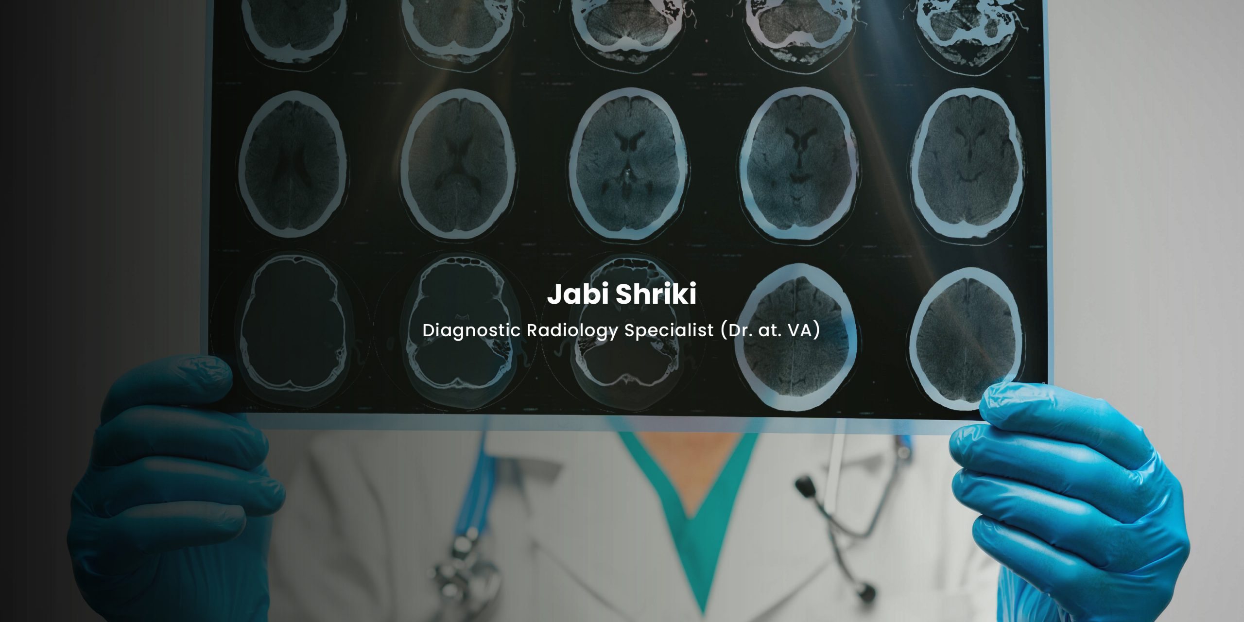
Radiology is critical in diagnosing, treating, and monitoring cardiovascular diseases. Advances in imaging technology have made it possible to detect heart conditions early and develop precise treatment strategies. By combining radiology with cardiology, healthcare professionals can gain detailed insights into the structure and function of the heart and blood vessels, significantly improving patient outcomes.
The evolution of radiology has paved the way for innovative approaches to cardiovascular disease management. With non-invasive techniques, radiologists can assess heart function without resorting to more intrusive procedures, allowing for a broader range of diagnostic options that benefit patients across all stages of care.
The Role of Non-Invasive Imaging Modalities
Non-invasive radiological techniques have transformed cardiovascular care by providing highly detailed images without surgical intervention. The most commonly used methods are echocardiography, cardiac MRI, and computed tomography (CT) scans. These modalities allow physicians to evaluate the heart’s anatomy, detect abnormalities, and assess the effectiveness of treatments.
Echocardiography, for instance, uses ultrasound waves to create detailed images of the heart in real-time, making it a valuable tool for diagnosing heart valve issues, cardiomyopathies, and congenital disabilities. Similarly, cardiac MRI offers a high-resolution view of the heart’s structures, including soft tissues, while providing essential information about blood flow and heart function. CT angiography, on the other hand, is instrumental in identifying blockages or abnormalities in the coronary arteries, aiding in diagnosing coronary artery disease.
Early Diagnosis and Prevention
Radiology has become vital in the early detection of cardiovascular diseases, which is crucial for preventing more severe health issues. Techniques such as coronary calcium scoring through CT scans help determine the risk of coronary artery disease by measuring the amount of calcium in the arterial walls. Identifying calcified plaque early enables physicians to implement preventive measures, reducing the risk of heart attacks or strokes.
By utilizing radiological imaging for early detection, healthcare providers can create personalized treatment plans focusing on managing risk factors. For instance, a patient with high calcium scores might be advised to adopt lifestyle changes and begin medication before more severe complications arise. This proactive approach is key to mitigating long-term cardiovascular risks.
Guiding Interventional Procedures
Radiology is also critical during interventional procedures, ensuring precise and effective interventions. Fluoroscopy, a live X-ray imaging technique, is often used to guide catheter-based treatments, such as angioplasty or stent placement, in patients with narrowed or blocked arteries. Fluoroscopy helps cardiologists safely navigate tools through the vascular system by providing real-time visualization of the blood vessels.
In more complex cases, hybrid procedures involving a combination of interventional cardiology and radiology are becoming increasingly common. For example, during transcatheter aortic valve replacement (TAVR), imaging ensures the new valve is accurately placed without needing open-heart surgery. These minimally invasive techniques reduce recovery times and complications, offering patients a less risky alternative to traditional surgery.
Monitoring Treatment Progress
The role of radiology in cardiovascular care extends beyond diagnosis and intervention—it is also a key component of treatment monitoring. After procedures such as angioplasty or bypass surgery, imaging techniques like echocardiography, MRI, and CT are used to assess how well the heart responds to treatment. This allows for early detection of complications and helps ensure the chosen therapies are effective.
Continuous monitoring through radiology is especially beneficial for patients with chronic cardiovascular conditions. For example, heart failure patients can undergo regular imaging to evaluate heart function and adjust their treatment plans as needed. This ongoing assessment is essential for managing long-term health outcomes and preventing further heart deterioration.
Emerging Trends in Cardiovascular Radiology
The field of radiology in cardiovascular disease management continues to evolve with new technologies and techniques. One promising development is using 3D imaging and artificial intelligence (AI) in interpreting radiological data. AI algorithms can analyze vast amounts of imaging data to detect patterns that may be missed by human eyes, potentially leading to earlier and more accurate diagnoses.
In addition, molecular imaging is gaining attention for its ability to visualize biological processes at the cellular level, offering new insights into the underlying causes of cardiovascular diseases. Tailoring treatments to the specific nature of the disease can lead to more targeted therapies and improve patient outcomes. These innovations can potentially transform how cardiovascular diseases are diagnosed and treated.
The Future of Radiology in Cardiovascular Care
Radiology’s contribution to cardiovascular disease management has revolutionized how conditions are diagnosed, treated, and monitored. From non-invasive imaging techniques to guiding complex interventional procedures, radiology provides essential tools that enhance patient care. With ongoing advancements in technology, including AI and molecular imaging, the future of cardiovascular radiology holds even greater promise for improving outcomes.
As radiology integrates with other medical fields, its role in personalized medicine will likely expand, offering tailored treatment plans and reducing the risk of complications. The synergy between radiology and cardiovascular care is indispensable, and its future developments will undoubtedly lead to more effective management strategies for patients with heart diseases.